Figure 6 from Femoral Hernia: A Review of the Clinical Anatomy and
Figure 6. Femoral hernia repair in clean operation. (a) The narrow side of the mesh is sutured to Cooper’s ligament; (b) The mesh is sutured to the iliopubic tract or shelving portion of the inguinal ligament; (c) The posterior wall of the inguinal canal is reinforced, as in Lichtenstein’s repair. - "Femoral Hernia: A Review of the Clinical Anatomy and Surgical Treatment"

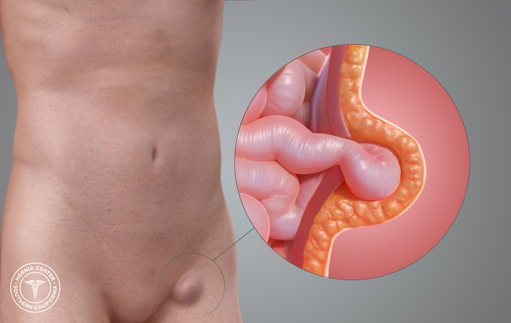
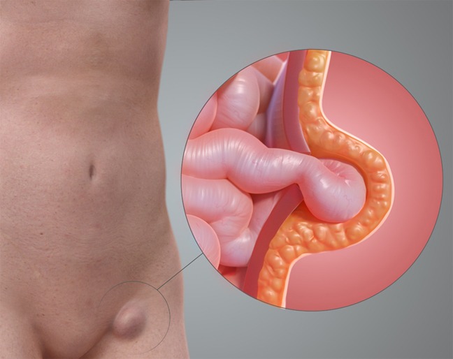
Femoral Hernia - A Review of Clinical Anatomy

Embryonic developmental process and clinical anatomy of the preperitoneal fascia and its clinical significance

Femoral Hernia - A Review of Clinical Anatomy

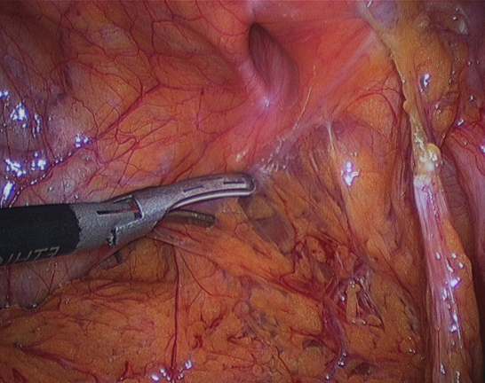
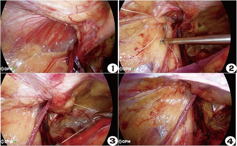
Laparoscopic view on a left sided femoral hernia. Arrows show the

Richter Hernia: Surgical Anatomy and Technique of Repair

Clinical Anatomy of the Groin: Posterior Laparoscopic Approach

Femoral hernia on herniography, Radiology Case
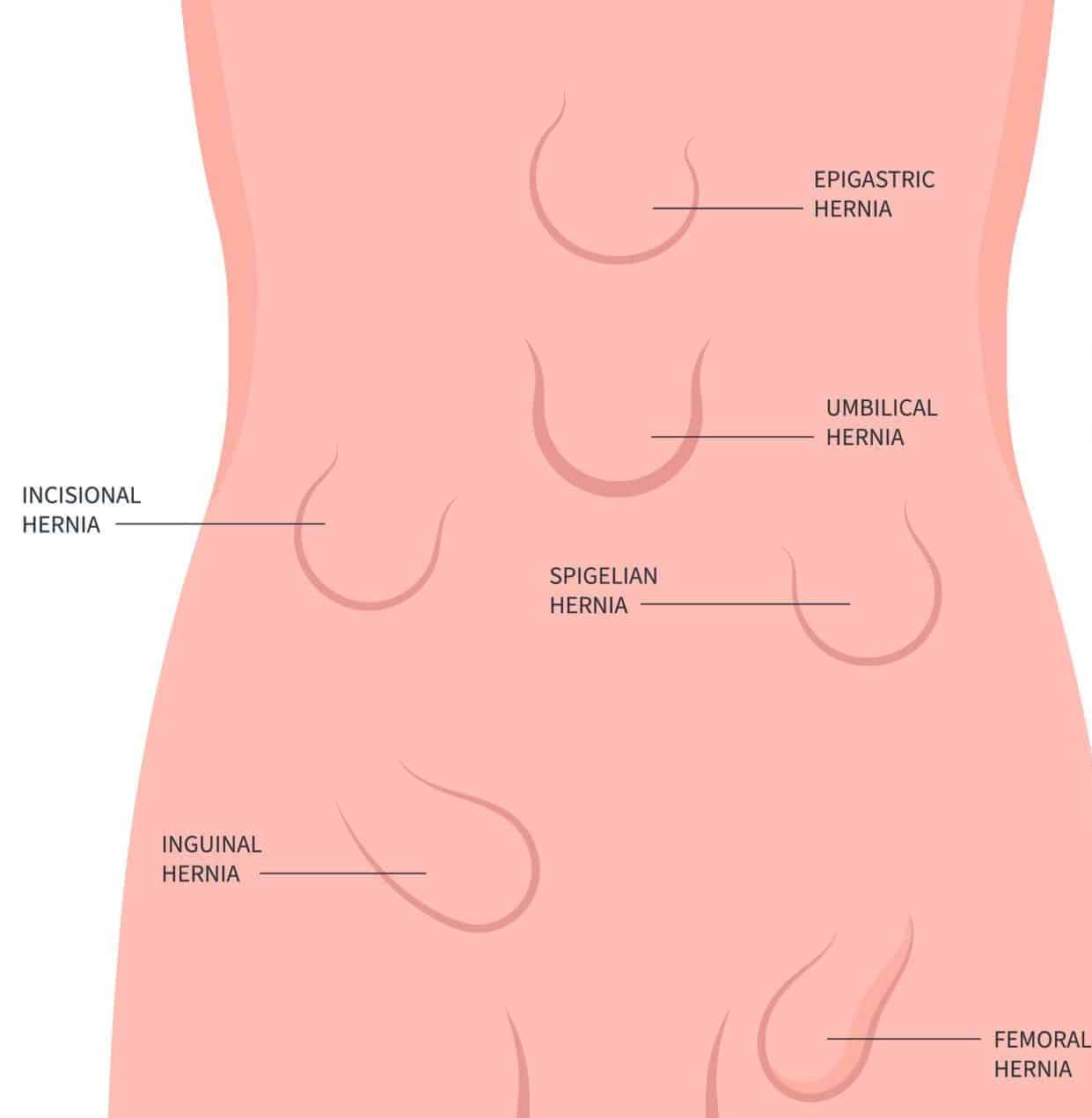
Abdominal Hernia - Epigastric - Spigelian - Obturator - TeachMeSurgery
Frontiers Clinical efficacy of laparoscopic closed hernia ring combined with a patch repair for Gilbert type III indirect inguinal hernia

Illustration Of A Femoral Hernia Art Print by John Bavosi - Fine Art America