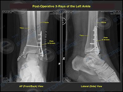
Left Ankle Fracture and Internal Fixation
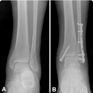
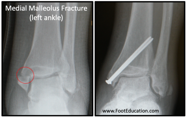
This exhibit features three radiological colorizations showing an ankle fracture and subsequent internal fixations. The first image depicts a fracture of the distal fibula, fracture of the distal tibia, and disruption of the ankle mortise. The second shows reduction of the fracture fragments with the placement of a fibular plate and multiple screws. Lastly, the third image illustrates fusion of the tibiofibular joint with a syndesmotic screw to reduce widening of the ankle mortise.

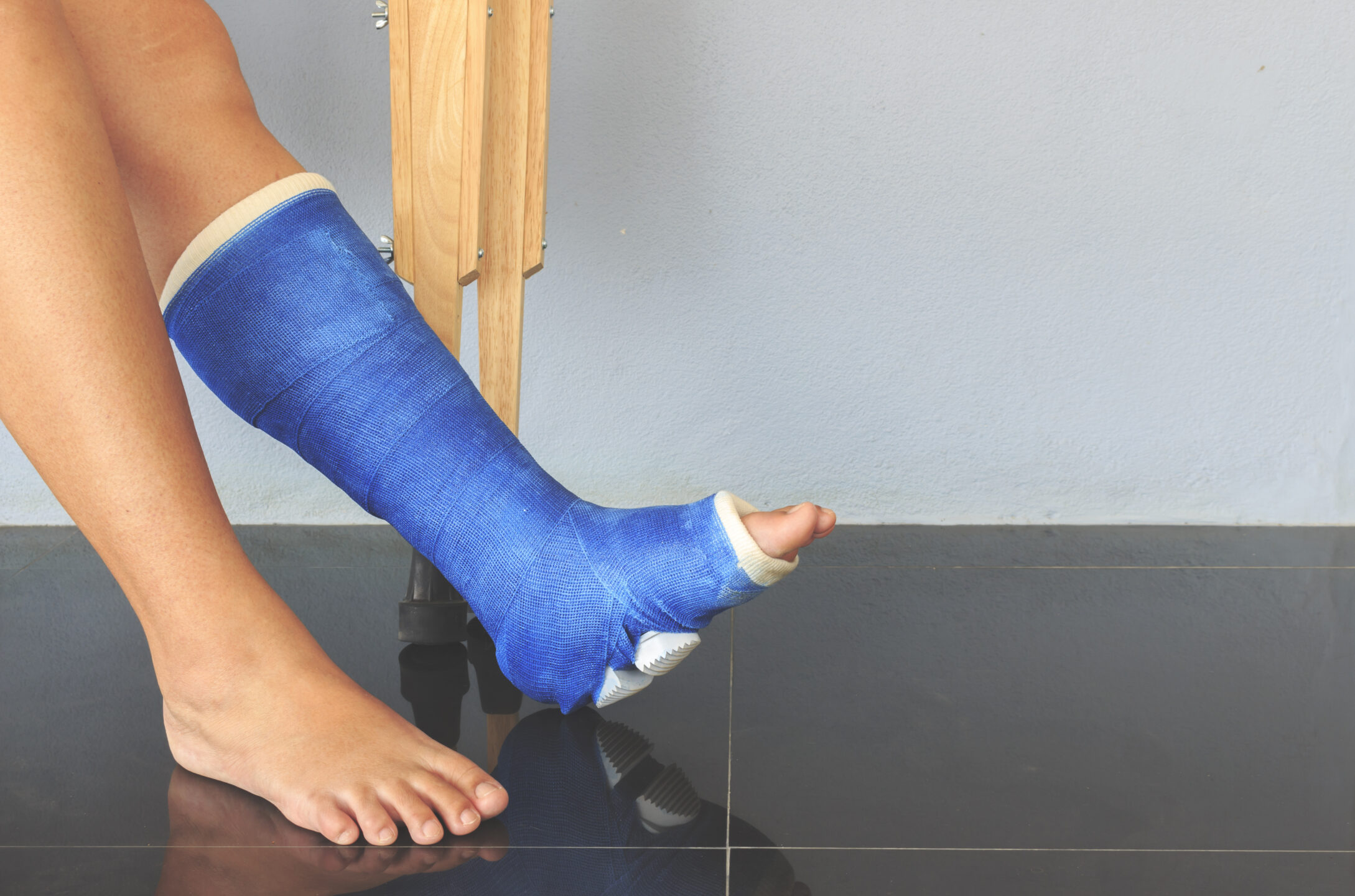
Early Weight-Bearing Following Ankle Fracture ORIF
Ankle Fracture ORIF - FootEducation
What Ankle Fracture Treatment is Right for You?
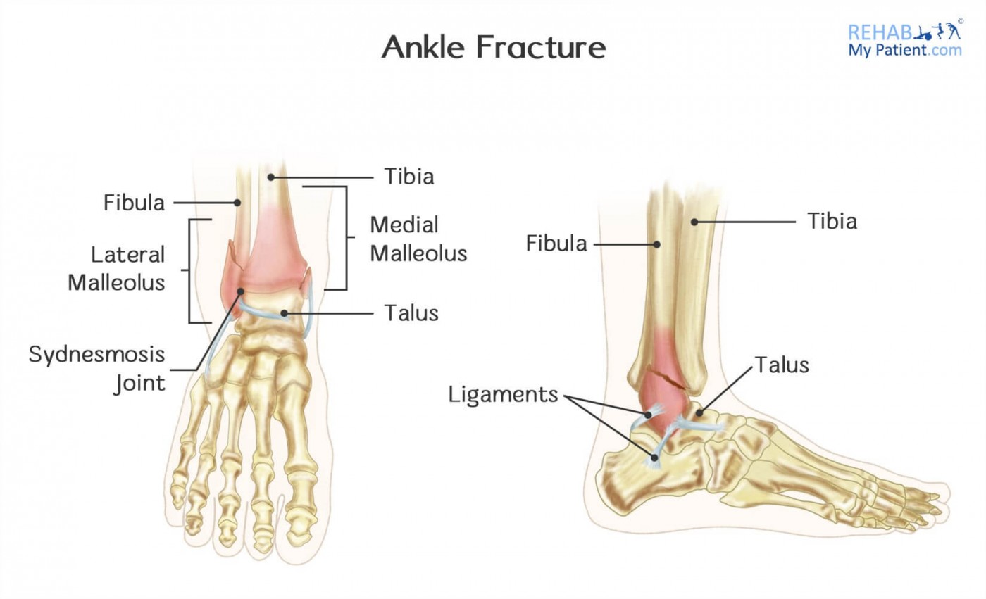
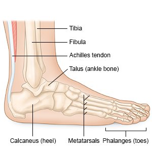
Ankle Fractures (Broken Ankle) - OrthoInfo - AAOS
ORIF of an Ankle Fracture - What You Need to Know

Early Weight-Bearing Following Ankle Fracture ORIF

Ankle Fracture Surgery Central Coast Orthopedic Medical Group

Medical Illustration Open Reduction Internal Fixation of Right
Accident Injuries: What is Open Reduction and Internal Fixation?

Ankle Joint Art Print By Sebastian Kaulitzki/science Photo, 45% OFF
Ankle Fractures (Broken Ankle) - OrthoInfo - AAOS