Diagnostics, Free Full-Text
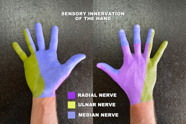
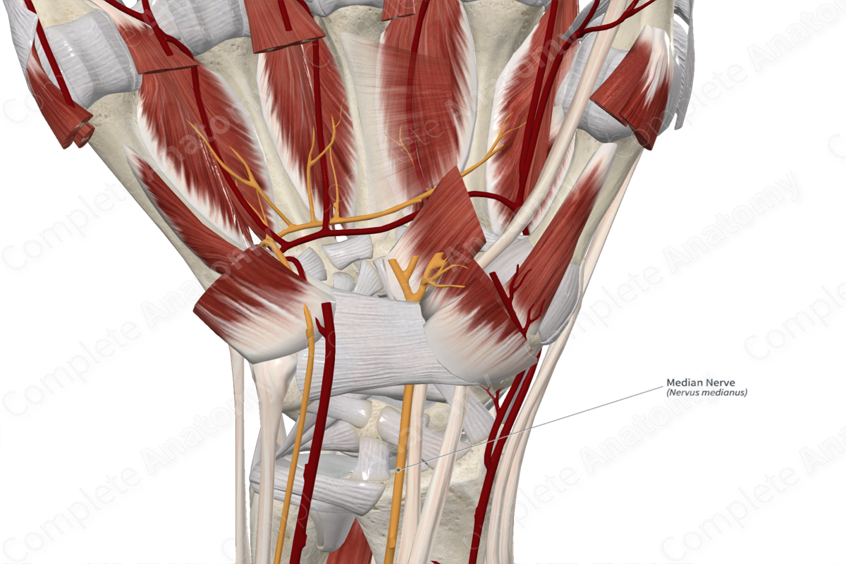
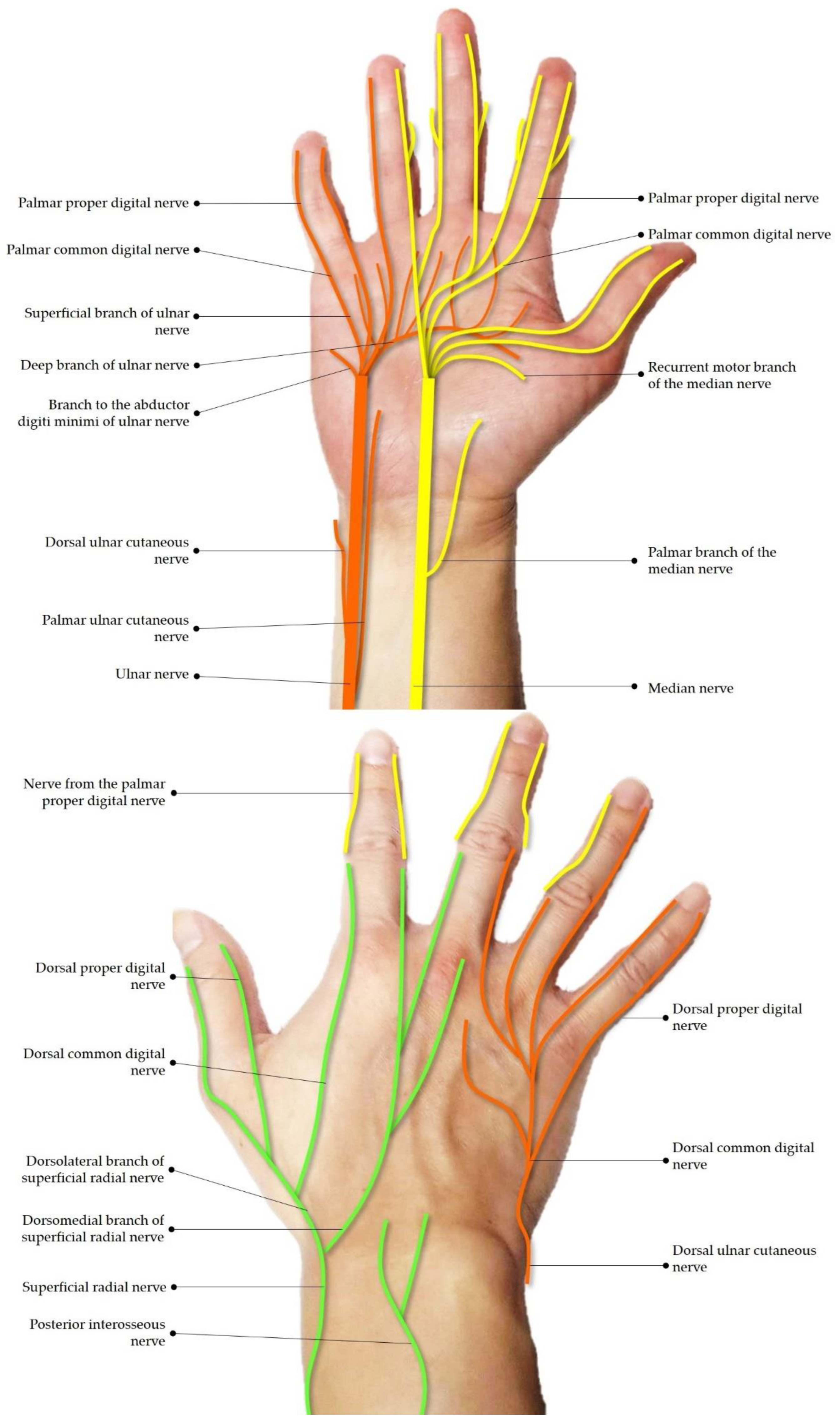
Ultrasound has emerged as a highly valuable tool in imaging peripheral nerve lesions in the wrist region, particularly for common pathologies such as carpal tunnel and Guyon’s canal syndromes. Extensive research has demonstrated nerve swelling proximal to the entrapment site, an unclear border, and flattening as features of nerve entrapments. However, there is a dearth of information regarding small or terminal nerves in the wrist and hand. This article aims to bridge this knowledge gap by providing a comprehensive overview concerning scanning techniques, pathology, and guided-injection methods for those nerve entrapments. The median nerve (main trunk, palmar cutaneous branch, and recurrent motor branch), ulnar nerve (main trunk, superficial branch, deep branch, palmar ulnar cutaneous branch, and dorsal ulnar cutaneous branch), superficial radial nerve, posterior interosseous nerve, palmar common/proper digital nerves, and dorsal common/proper digital nerves are elaborated in this review. A series of ultrasound images are used to illustrate these techniques in detail. Finally, sonographic findings complement electrodiagnostic studies, providing better insight into understanding the whole clinical scenario, while ultrasound-guided interventions are safe and effective for treating relevant nerve pathologies.
Diagnostics, Free Full-Text
Diagnostics, Free Full-Text
Diagnostics, Free Full-Text

Husnain Sherazi on LinkedIn: #teamwork #research #uclautism #autism

Diagnostics, Free Full-Text
Diagnostics, Free Full-Text
FIGURE. Diagnosis and treatment in 284 consecutive patients with
💕👉 {U=9} 2024 cumshot anatomy

Diagnostics, Free Full-Text
Diagnostics, Free Full-Text

